Male Pelvis Anatomy Front View Labeled Diagram Ilustrasi Stok Biology Diagrams
Male Pelvis Anatomy Front View Labeled Diagram Ilustrasi Stok Biology Diagrams This e-Anatomy module contains seventy-nine illustrations dedicated to the anatomy of the male pelvis and genital system. These fully annotated anatomical illustrations are presented as a comprehensive atlas of the male reproductive system, specifically designed for medical students, residents and healthcare professionals. Material and methods
Learn about the anatomy and physiology of the male pelvis with a free printable model and a virtual 3D pelvis app. This course is funded by Plus online learning and requires registration to access.
22: The Reproductive System (Male) Biology Diagrams
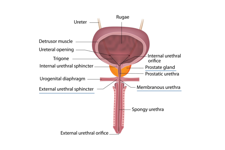
Learn about the anatomy of the male reproductive system, including the seminal vesicles, the bulbourethral glands and the urethra. The pelvis is the lower portion of the trunk that connects the axial skeleton to the lower limbs and contains many organs. Learn about the anatomy of the male pelvis, including prostate, bladder, genital organs, rectum and more. See MRI images and labels of the pelvic structures and their zones, regions and fascia.
Learn about the bony, muscular and soft tissue structures of the pelvis and perineum, and how they differ between males and females. Find out the functions, innervation and blood supply of the pelvic organs and perineal membrane.
Anatomy of the male reproductive organs of the pelvis Biology Diagrams
Learn about the anatomy and function of the ductus deferens, seminal vesicles, ejaculatory ducts, and prostate in the male pelvis. Watch video, view diagrams, and test your knowledge with quizzes. 22.6: MODELS- Male Hemi-Pelvis and Torso This page provides a detailed overview of the male reproductive system's anatomy and histology, covering structures like the scrotum, testes, ejaculatory duct, urethra, and penis, along with components such as the dartos muscle and seminal vesicles. This MRI male pelvis axial cross sectional anatomy tool is absolutely free to use. Use the mouse scroll wheel to move the images up and down, or alternatively, use the tiny arrows (→) on both sides of the image to navigate through the images. For a more detailed view, double-click the image to view it in full screen, and use the menu in the